Radiologia Brasileira - Publicação Científica Oficial do Colégio Brasileiro de Radiologia
AMB - Associação Médica Brasileira CNA - Comissão Nacional de Acreditação
 Vol. 48 nº 2 - Mar. / Apr. of 2015
|
LETTER TO THE EDITOR
|
|
Catamenial pneumothorax |
|
|
Autho(rs): Brainner Campos Barbosa1; Edson Marchiori2; Gláucia Maria Ribeiro Zanetti2; Jorge Luiz Barillo3 |
|
|
Dear Editor,
A previously healthy 29-year-old woman presented at the emergency service complaining of sudden onset dyspnea. At physical examination the vesicular murmur was absent in the entire right hemithorax. Chest radiography demonstrated the presence of pneumothorax at right (Figure 1) and chest computed tomography (CT) did not demonstrate any other alteration besides the already mentioned pneumothorax. Thoracotomy with underwater seal chest drainage was performed. As the pneumothorax presentation coincided with the patient's menstrual period, pelvic ultrasonography was performed and identified an image compatible with endometrioma in the left ovary. In three months, the patient evolved with a new spontaneous pneumothorax at right, and a pig-tail drainage tube was inserted. Later, thoracocoscopy was performed, and endometriotic foci were identified and resected (Figure 2). The chest wall was repaired with a Marlex mesh. After three months, the patient remains asymptomatic. 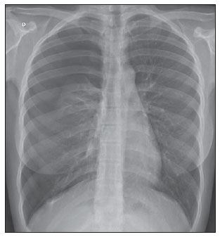 Figure 1. Chest radiography, posterior view showing pneumothorax at right. 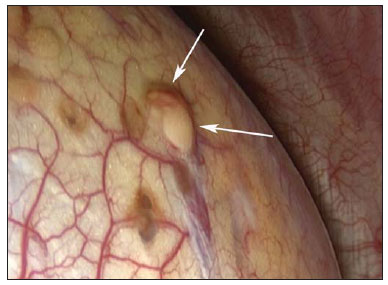 Figure 2. Videothoracoscopy revealing the presence of endometriotic foci (arrows) on the pleural surface. Imaging evaluation of the chest has been subject of a range of recent publications in the Brazilian radiological literature(1-10). Thoracic endometriosis is the presence of endometrial tissue in the lung parenchyma or in the pleural cavity, and manifests clinically by hemoptysis, pneumothorax or hemothorax, occurring in conjunction with menstrual periods(11-13). Frequently, it affects women in childbearing age, with incidence peak between the third and fourth decades of life(14). Thoracic endometrial implants generally occur in the pleural cavity and, less frequently, in the lung parenchyma(14). Pleural endometriosis is an entity whose course is generally benign, occurring most frequently at right, possibly due to congenital defects in the right diaphragmatic dome and to continuous flow of fluid from the pelvis into the right upper quadrant of the abdomen(15). Typically, thoracic endometrial implants occur concurrently with periodical symptoms (from one day before to the first two days of menstruation)(16). The clinical presentation depends on the site of involvement, namely, catamenial pneumothorax or hemothorax in cases of pleural implants; and catamenial hemoptysis or asymptomatic pulmonary nodules in cases of implants in the pulmonary parenchyma. Histologically, the presence of endometrial tissue is identified in the lungs and/or pleura. Cytology reveals the presence of endometrial cells in the pleural fluid, in pulmonary nodules/masses aspirates, or in bronchial lavage fluid. Imaging studies include principally chest radiography and CT, which can demonstrate pneumothorax, hydropneumothorax or pleural nodular lesions. Magnetic resonance imaging has increasingly gained relevance since, besides differentiating parenchymal from pleural lesions, this method presents a better spatial resolution and, if performed during the menstrual period, it can identify glandular tissue in the affected site (hyperintense foci on T2-weighted images)(17-19). The treatment has two main pillars: the conservative treatment, based on hormone replacement to prevent recurrence of pneumothorax and hemothorax; and the surgical treatment that is indicated in cases of hormone therapy failure, severe treatment side effects, recurrence after treatment interruption, or if the patient wants to become pregnant(20). Therefore, one may conclude that catamenial pneumothorax should be suspected in the presence of clinical signs coinciding with the menstrual period, and that imaging studies can confirm the diagnosis. The treatment may be either surgical or medical, and should be appropriately indicated to avoid disease recurrence. REFERENCES 1. Zanetti G, Nobre LF, Mançano AD, et al. Paracoccidioidomicose pulmonar. Radiol Bras. 2014;47(1):xi-xiii. 2. Fernandes MC, Zanetti G, Hochhegger B, et al. Pneumonia por Rhodococcus equi em paciente com SIDA. Radiol Bras. 2014;47(3):xi-xiii. 3. Amorim VB, Rodrigues RS, Barreto MM, et al. Achados na tomografia computadorizada em pacientes com infecção pulmonar pelo vírus influenza A (H1N1). Radiol Bras. 2013;46:299-306. 4. Eifer DA, Arsego FV, Torres FS. Atresia unilateral das veias pulmonares: avaliação por tomografia computadorizada. Radiol Bras. 2013;46:376-8. 5. Souza VF, Chaves RT, Balieiro VS, et al. Avaliação qualitativa e quantitativa da densidade pulmonar em paciente com polimiosite e fibrose pulmonar. Radiol Bras. 2013;46(3):ix-x. 6. Marcos L, Bichinho GL, Panizzi EA, et al. Classificação da doença pulmonar obstrutiva crônica pela radiografia do tórax. Radiol Bras. 2013;46:327-32. 7. Amoedo MK, Souza LVS, Souza AS, et al. Enfisema intersticial pulmonar: relato de caso e revisão da literatura. Radiol Bras. 2013;46:317-9. 8. Zanetti G, Nobre LF, Mançano AD, et al. Sinal do halo invertido com paredes nodulares causado por tuberculose pulmonar, confirmada por cultura do escarro. Radiol Bras. 2013;46(6):ix-x. 9. Koenigkam-Santos M, Paula WD, Gompelmann D, et al. Endobronchial valves in severe emphysematous patients: CT evaluation of lung fissures completeness, treatment radiological response and quantitative emphysema analysis. Radiol Bras. 2013;46:15-22. 10. Silva JLP. O trinômio vírus-droga-hospedeiro na caracterização tomográfica da infecção pulmonar por influenza A (H1N1) - uma visão clínico-radiológico-patológica. Radiol Bras. 2013;46(5):vii-ix. 11. Alifano M, Vénissac N, Mouroux J. Recurrent pneumothorax associated with thoracic endometriosis. Surg Endosc. 2000;14:680. 12. Attaran S, Bille A, Karenovics W, et al. Videothoracoscopic repair of diaphragm and pleurectomy/abrasion in patients with catamenial pneumothorax: a 9-year experience. Chest. 2013;143:1066-9. 13. Bagan P, Berna P, Assouad J, et al. Value of cancer antigen 125 for diagnosis of pleural endometriosis in females with recurrent pneumothorax. Eur Respir J. 2008;31:140-2. 14. Costa F, Matos F. Endometriose torácica. Rev Port Pneumol. 2008;XIV:427-35. 15. Cassina PC, Hauser M, Kacl G, et al. Catamenial hemoptysis. Diagnosis with MRI. Chest. 1997;111:1447-50. 16. Yu Z, Fleischman JK, Rahman HM, et al. Catamenial hemoptysis and pulmonary endometriosis: a case report. Mount Sinai J Med. 2002;69:261-3. 17. Marchiori E, Zanetti G, Rafful P, et al. Pleural endometriosis and recurrent pneumothorax: the role of magnetic resonance imaging. Ann Thorac Surg. 2012;93:696-7. 18. Coutinho A Jr, Bittencourt LK, Pires CE, et al. MR imaging in deep pelvic endometriosis: a pictorial essay. Radiographics. 2011;31:549-67. 19. Marchiori E, Zanetti G, Rodrigues RS, et al. Pleural endometriosis: findings on magnetic resonance imaging. J Bras Pneumol. 2012;38:797-802. 20. Alifano M, Roth T, Broët SC, et al. Catamenial pneumothorax: a prospective study. Chest. 2003;124:1004-8. 1. Hospital Santa Teresa, Petrópolis, RJ, Brazil 2. Universidade Federal do Rio de Janeiro (UFRJ), Rio de Janeiro, RJ, Brazil 3. Universidade Federal Fluminense (UFF), Niterói, RJ, Brazil Mailing Address: Dr. Brainner Campos Barbosa Rua das Laranjeiras, 371, ap. 303, Edifício Marco Luiz, Laranjeiras Rio de Janeiro, RJ, Brazil, 22240-004 E-mail: brainnerc@gmail.com |
|
GN1© Copyright 2024 - All rights reserved to Colégio Brasileiro de Radiologia e Diagnóstico por Imagem
Av. Paulista, 37 - 7° andar - Conj. 71 - CEP 01311-902 - São Paulo - SP - Brazil - Phone: (11) 3372-4544 - Fax: (11) 3372-4554
Av. Paulista, 37 - 7° andar - Conj. 71 - CEP 01311-902 - São Paulo - SP - Brazil - Phone: (11) 3372-4544 - Fax: (11) 3372-4554