Radiologia Brasileira - Publicação Científica Oficial do Colégio Brasileiro de Radiologia
AMB - Associação Médica Brasileira CNA - Comissão Nacional de Acreditação
 Vol. 50 nº 2 - Mar. / Apr. of 2017
|
LETTER TO THE EDITOR
|
|
What radiologists need to know about 3D printing and its main applications in musculoskeletal imaging |
|
|
Autho(rs): Francisco Abaeté Chagas-Neto1; Francisco Coracy Carneiro Monteiro2; Eduardo Lima da Rocha3; Everaldo Gregio-Junior4; Marcello Henrique Nogueira-Barbosa4 |
|
|
Dear Editor,
The utility of the various imaging methods in the evaluation and diagnosis of musculoskeletal disorders is well established, and those methods play a fundamental role in the planning of the different treatments, be they conservative or surgical, providing images that can be manipulated through specific software to create three-dimensional (3D) reconstructions. To date, however, such 3D reconstructions have been made available only as digital files, as images on radiographic films, or as prints on paper. These traditional forms of image documentation do not always allow surgeons to have a real in-depth sensory notion and knowledge of the 3D anatomical relationships in the planning of different types of surgical procedures. Recently, 3D printing has come to be used with increasingly frequency to obtain a more realistic and more accurate analysis by creating 3D models(13). What really is 3D printing? The definition of 3D printing, also known as rapid prototyping, is the use of a set of methods to create solid three-dimensional objects (models or prototypes) from the data contained in digital files. There are different forms of 3D printing, one of the most popular being the additive processing technique, in which the object is created layer by layer through successive depositions of a highly resistant plastic polymer. How does 3D printing work? It all begins with the development of the 3D digital file. The file is obtained through the acquisition of sectional images through the use of magnetic resonance imaging, computed tomography, or even (3D or 4D) ultrasound. The digital file is then analyzed and processed with computer-aided design (CAD) software, according to what is required in each situation. After developing the 3D digital file, the CAD modeling software divides the prototype into hundreds or thousands of thin horizontal layers, thus preparing the file for printing. The digital file can then be loaded into a 3D printer for printing. Is 3D printing already a reality in clinical practice or only in experimental research? In several countries, it is already a part of the clinical routine, having been shown to have a great impact on the precision and safety of surgical procedures(25). There has been rapid growth in the number of potential applications of 3D printing in medicine, which has already been used in several situations, even in Brazil(68). We illustrate, as an example, a case in which 3D printing was employed at our facility for the preoperative planning of the surgical treatment of an osteolytic lesion in the mandible (Figure 1). 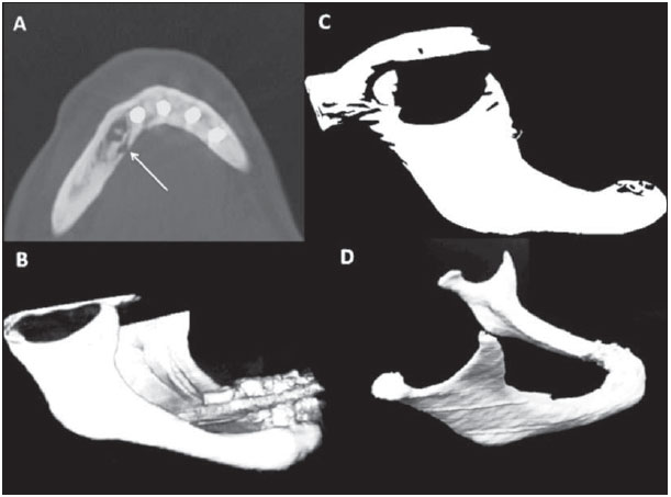 Figure 1. A: Axial computed tomography slice showing an osteolytic lesion in the right mandible (arrow). B: 3D volume-rendering computed tomography reconstruction of the mandible. C: 3D digital file, containing images of the mandible, being analyzed and processed in CAD software. D: Final aspect of the mandible prototype printed in 3D to mold the osteosynthesis material before surgery. What are the applications of 3D printing in musculoskeletal imaging? Among its many potential applications in clinical practice and in the teaching of medicine, we emphasize in this article the use of the technique in musculoskeletal imaging. We highlight its application in the preoperative planning of complex surgical procedures, which require high precision, such as those employed in the treatment of spinal deformities and complex fractures, as well as in the creation of models of orthotics and prostheses tailored to the anatomy and needs of each patient(19). We believe it to be inevitable that, in the coming years, there will be growth in the application of the 3D printing technique in the field of medicine as a whole, especially in the area of musculoskeletal imaging. The incorporation of this new technique will allow the optimization of protocols promoting good practices, offering greater effectiveness to the professionals involved and allowing better results, with potentially greater safety for patients. REFERENCES 1. Jones DB, Sung R, Weinberg C, et al. Three-dimensional modeling may improve surgical education and clinical practice. Surg Innov. 2016; 23:18995. 2. Wu C, Tan L, Lin X, et al. Clinical application of individualized reference model of sagittal curves by three-dimensional printing technique and computed-aided navigation system for lumbar spondylolystesis. Zhongguo Xiu Fu Chong Jian Wai Ke Za Zhi. 2015;29:73440. 3. Chen X, Zhang G, Lin H, et al. Digital design of standard parts database for proximal tibia fractures treated with plating via three-dimensional printing. Zhongguo Xiu Fu Chong Jian Wai Ke Za Zhi. 2015;29:70411. 4. Mowry SE, Jammal H, Myer C 4th, et al. A novel temporal bone simulation model using 3D printing techniques. Otol Neurotol. 2015;36:15625. 5. Xu N, Wei F, Liu X, et al. Reconstruction of the upper cervical spine using a personalized 3D-printed vertebral body in an adolescent with Ewing sarcoma. Spine (Phila Pa 1976). 2016;41:E504. 6. Werner H, Rolo LC, Araujo Júnior E, et al. Manufacturing models of fetal malformations built from 3-dimensional ultrasound, magnetic resonance imaging, and computed tomography scan data. Ultrasound Q. 2014;30:6975. 7. Werner Jr H, Santos JL, Belmonte S, et al. Applicability of three-dimensional imaging techniques in fetal medicine. Radiol Bras. 2016;49:2817. 8. Araujo Júnior E. Three-dimensional ultrasound in fetal medicine after 25 years in clinical practice: many advances and some questions. Radiol Bras. 2016;49(5):vvi. 9. AbouHashem Y, Dayal M, Savanah S, et al. The application of 3D printing in anatomy education. Med Educ Online. 2015;20:29847. 1. Centro Universitário Christus (Unichristus) e Hospital Antônio Prudente, Fortaleza, CE, Brazil 2. Hospital Albert Sabin, Fortaleza, CE, Brazil 3. Hospital Antônio Prudente, Fortaleza, CE, Brazil 4. Faculdade de Medicina de Ribeirão Preto da Universidade de São Paulo (FMRP-USP), Ribeirão Preto, SP, Brazil Mailing address: Dr. Francisco Abaeté Chagas-Neto Rua João Adolfo Gurgel, 133, Coco Fortaleza, CE, Brazil, 60192-345 E-mail: fabaeteneto@gmail.com |
|
GN1© Copyright 2024 - All rights reserved to Colégio Brasileiro de Radiologia e Diagnóstico por Imagem
Av. Paulista, 37 - 7° andar - Conj. 71 - CEP 01311-902 - São Paulo - SP - Brazil - Phone: (11) 3372-4544 - Fax: (11) 3372-4554
Av. Paulista, 37 - 7° andar - Conj. 71 - CEP 01311-902 - São Paulo - SP - Brazil - Phone: (11) 3372-4544 - Fax: (11) 3372-4554