Radiologia Brasileira - Publicação Científica Oficial do Colégio Brasileiro de Radiologia
AMB - Associação Médica Brasileira CNA - Comissão Nacional de Acreditação
 Vol. 51 nº 2 - Mar. / Apr. of 2018
|
NEWS IN RADIOLOGY
|
|
Ultrasound-guided biopsy of breast calcifications using a new image processing technique: initial experience |
|
|
Autho(rs): Almir Galvão Vieira Bitencourt1; Luciana Graziano2; Camila Souza Guatelli2; Maria Luiza Lima Albuquerque3; Elvira Ferreira Marques3 |
|
|
Keywords: Ultrasonography, mammary/methods; Breast diseases/diagnosis; Biopsy, needle/methods. |
|
|
Abstract: INTRODUCTION
Mammography is the method of choice to guide biopsies of suspicious breast calcifications(1,2). However, despite the use of alternative approaches(2,3), stereotactic biopsy is not technically achievable in all cases. Ultrasound has been proposed as an alternative to stereotactic biopsy in selected patients with suspicious breast calcifications(4,5). However, conventional gray-scale examination limits the identification of calcifications, especially those located outside of a mass or duct, due to the lack of contrast with the normal breast parenchyma. Recent technical advances have improved the detection of calcifications by ultrasound(68). The aim of this paper is to describe the use of a new ultrasound imaging processing technique to identify and guide biopsies of suspicious breast calcifications detected on mammography. MATERIALS AND METHODS From June 2014 through June 2016, 13 patients with suspicious grouped breast calcifications (< 2 cm in extent) that could not be submitted to stereotactic biopsy were submitted to ultrasound-guided biopsy after the identification of calcifications using a new imaging processing technique (MicroPure; Toshiba Medical Systems, Tokyo, Japan). MicroPure is an image processing function designed to enhance the visualization of microcalcifications by ultrasound. It combines nonlinear images and suppression techniques to highlight suspicious calcifications as white spots on a blue background. During ultrasound examination, gray-scale and MicroPure images are displayed side-by-side on the screen. All of the patients gave written informed consent prior to the procedure. Three radiologists with experience in breast imaging performed the ultrasound examinations and biopsies. Mammography images were analyzed to evaluate the distribution and topographic location of the calcifications. Ultrasound was then performed on an Aplio 500 system (Toshiba Medical Systems, Tokyo, Japan) with a high-frequency linear transducer. After identification of the suspicious calcifications on ultrasound, 12-gauge core needle biopsy was performed. Specimen X-ray images were obtained to confirm that the calcifications were present (Figure 1). 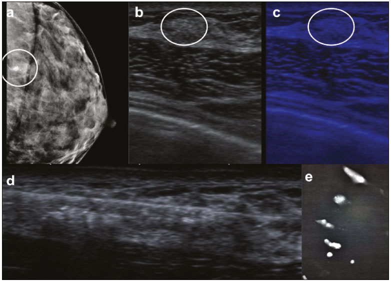 Figure 1. A 36-year-old asymptomatic woman at high risk for breast cancer. a: Mammography showing grouped amorphous calcifications in the left breast (circle), near the chest wall, which precluded the use of stereotactic biopsy. b,c: Ultrasound showing the calcifications (circle) on gray-scale imaging (b) and MicroPure imaging (c). d: Ultrasound-guided 12-gauge core needle biopsy, the pathological analysis of which was consistent with atypical ductal hyperplasia. e: X-ray image of the specimens, confirming the presence of calcifications. RESULTS In all 13 of the cases evaluated, stereotactic biopsy could not be performed, because of the location of the lesion in five cases (the lesion being near the skin in one, near the chest wall in one, and near breast implants in three); because of insufficient thickness of the compressed breast in three; and because of clinical conditions in five (one obese patient, one patient who refused to undergo the procedure, one patient with abdominal sarcoma, and two elderly patients with comorbidities that prevented them from assuming the prone position). Suspicious calcifications were identified using ultrasound, and the biopsy was successfully performed in all cases, without complications. The main technical difficulty was to identify suspicious calcifications after removal of the first sample, due to the presence of gas and edema/hematoma at the biopsy site. However, that did not impede the removal of calcifications in the additional samples. Chest X-ray revealed microcalcifications in all specimens. Table 1 summarizes the characteristics of the lesion, pathological results, and outcomes. 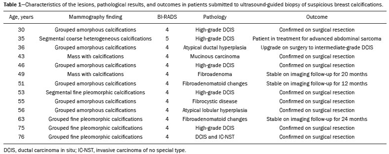 DISCUSSION The technique described here improves the capacity of ultrasound to detect calcifications and increases the confidence of radiologists in their ability to perform ultrasound-guided biopsy in selected cases, when there are technical challenges for stereotactic biopsy due to the lesion location or clinical characteristics of the patients. There have been few reports on the clinical utility of the MicroPure technique in locating and evaluating breast calcifications(7,8). Park et al.(6) recently used the MicroPure ultrasound function to guide biopsies of suspicious calcifications in nine patients. As in the present study, ultrasound biopsy was successfully performed in all patients. Further large-scale studies are needed in order to assess the potential contribution of this new technique and to compare its performance with that of stereotactic biopsy in the diagnosis of suspicious calcifications. In conclusion, although mammography remains the gold standard for the histological evaluation of suspicious breast calcifications, ultrasound using innovative imaging processing techniques can be an alternative means of guiding biopsies, thus avoiding unnecessary surgery, in selected patients. REFERENCES 1. Sickles EA, D''Orsi CJ, Bassett LW, et al. ACR BI-RADS® Mammography. In: ACR BI-RADS® Atlas, Breast Imaging Reporting and Data System. Reston, VA: American College of Radiology; 2013. 2. Mahoney MC, Newell MS. Breast intervention: how I do it. Radiology. 2013;268:1224. 3. Huang ML, Adrada BE, Candelaria R, et al. Stereotactic breast biopsy: pitfalls and pearls. Tech Vasc Interv Radiol. 2014;17:329. 4. Kim HS, Kim MJ, Kim EK, et al. US-guided vacuum-assisted biopsy of microcalcifications in breast lesions and long-term follow-up results. Korean J Radiol. 2008;9:5039. 5. Soo MS, Baker JA, Rosen EL, et al. Sonographically guided biopsy of suspicious microcalcifications of the breast: a pilot study. AJR Am J Roentgenol. 2002;178:100715. 6. Park AY, Seo BK, Cho KR, et al. The utility of MicroPure ultrasound technique in assessing grouped microcalcifications without a mass on mammography. J Breast Cancer. 2016;19:836. 7. Tan R, Xiao Y, Tang Q, et al. The diagnostic value of Micropure imaging in breast suspicious microcalcification. Acad Radiol. 2015;22: 133843. 8. Machado P, Eisenbrey JR, Cavanaugh B, et al. Microcalcifications versus artifacts: initial evaluation of a new ultrasound image processing technique to identify breast microcalcifications in a screening population. Ultrasound Med Biol. 2014;40:23214. 1. MD, PhD, Radiologist at A.C.Camargo Cancer Center, São Paulo, SP, Brazil 2. MD, MSc, Radiologist at A.C.Camargo Cancer Center, São Paulo, SP, Brazil 3. MD, Radiologist at A.C.Camargo Cancer Center, São Paulo, SP, Brazil Study conducted in the Department of Imaging of the A.C.Camargo Cancer Center, São Paulo, SP, Brazil. Mailing address: Dr. Almir Galvão Vieira Bitencourt A.C.Camargo Cancer Center Departamento de Imagem Rua Professor Antônio Prudente, 211, Liberdade São Paulo, SP, Brazil, 01509-010 E-mail: almirgvb@yahoo.com.br Received March 26, 2017. Accepted after revision May 8, 2017. |
|
Av. Paulista, 37 - 7° andar - Conj. 71 - CEP 01311-902 - São Paulo - SP - Brazil - Phone: (11) 3372-4544 - Fax: (11) 3372-4554